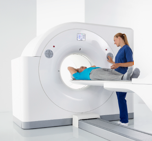
PET Scan

A cardiac PET/CT exam, also known as Cardiac Positron Emission Tomography/Computed Tomography, is a specialized imaging test used to evaluate the blood flow and function of your heart. A cardiac PET scan can provide information other imaging tests cannot and the test can find problems earlier than other tests can.
This scan is an accurate, noninvasive test that creates images of your heart. A healthcare provider can use these images to make decisions about the next steps in your heart care. They can judge how healthy your heart muscle is and decide how to treat it. A cardiac PET scan uses a small amount of radiation.
A cardiac PET (positron emission tomography) scan creates images of your heart using a scanning machine and an injection of radioactive tracers. The test’s radioactive tracers release energy. Depending on the specific type of tracer and the conditions under which a healthcare provider injects it, the pattern with which the tracer lights up your heart can give providers information about how healthy your heart is.
This highly effective imaging tool is used to diagnose cardiac disease with higher accuracy and lower radiation exposure than traditional stress testing. PET/CT imaging provides diagnostic, excellent quality, and the ability to detect disease before symptoms are present.
This non-invasive test is the gold standard in cardiac perfusion imaging, and we couldn’t be prouder to offer it.
When is a cardiac PET/CT scan performed?
A provider performs a PET/CT scan of your heart when they need to know:
- Which areas in your heart don’t have good blood flow.
- If you have coronary artery disease.
- If you have diseased heart muscle.
- If you have an infection in your heart.
- If you have cardiac sarcoidosis.
- How much heart damage you have after a heart attack.
- How well your cardiac treatment plan is working.
- How healthy your heart is before a procedure.
